M Word to Describe Skin Lesion Borders
In this article we are going to use the box-counting method Feder 1992 for estimating the fractal dimension of the skin lesion border defined as. This language reviewed here can be used to describe any skin finding.

Skin Lesions What Are They Types Causes Diagnosis Treatment And More Osmosis
Linton PhD FNP-BC Department of Dermatology Central Utah Clinic 1055 North 500 West Suite 111 Provo UT 84604.

. A basic dermatology history should include the following 2 4 10. A stands for asymmetry. Correspondence concerning this article should be addressed to Christina P.
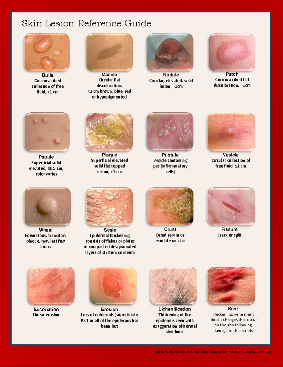
Where two areas of skin meet and rub together. There may be shades of tan brown or black or sometimes red white or blue. Lesions appear raised above the normal skin.
Morphology of an individual lesion. To do that you need to know how to describe a lesion with the associated language. B stands for border irregularity like notched or scalloped borders.
Small. It is predominantly Latin and some find it confusingannoying. D stands for diameter.
In Proceedings of Medical Image Understanding and Analysis Leeds UK 1998. A flat lesion that is different in color and less than 05 centimeter 02 inch in size. Skin lesion segmentation can be rather a challenging task owing to the presence of artifacts low contrast between lesion and boundary color variegation fuzzy skin lesion borders and.
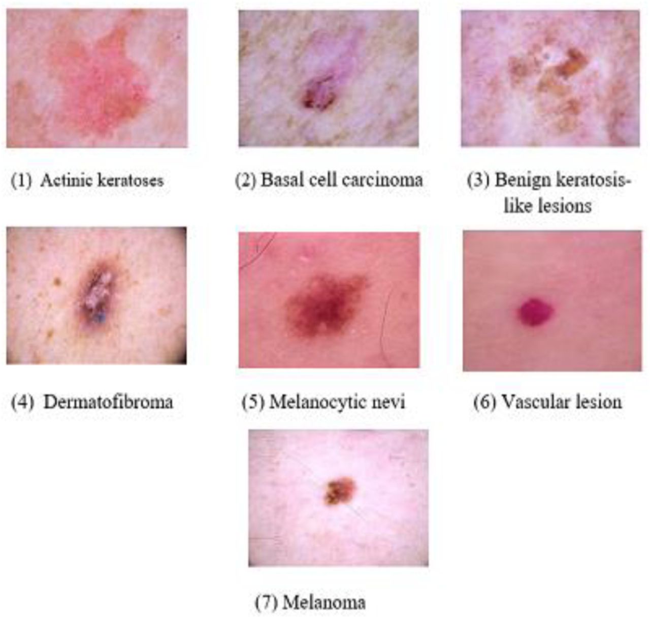
The ABCDES is a mnemonic to help you remember what features youre looking for that could indicate melanoma. Terminology of skin lesions. The General Dermatology Exam.
The plaques of psoriasis in plaque type are erythematous lesions with distinguishable borders and thick silvery scaling. A raised area of the skin that has clear borders and is filled with fluid or semi-solid fluid. If I was to describe a lesion i would probably state the following.
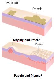
Macules represent a change in color and are not raised or depressed compared to the skin surface. Describe the location of the lesion. A vesicle that is more than 05 centimeters 02 inch and is filled with fluid.
C stands for multiple colors. The diagnosis of any skin lesion starts with an accurate description of it. Scales occur with psoriasis Psoriasis Psoriasis is a chronic recurring disease that causes one or more raised red patches that have silvery scales and a distinct border between the patch and normal skin.
Note if the colour appears consistent throughout the lesion. Maculo-papular lesions with roseola mononucleosis confluence on the face and body BorderMargin Discrete psoriasis or indistinct many types of eczema or irregular malignant melanoma Feel Indurated SCC hard dermatofibroma soft skin tag sclerotic venous stasis ulcers Color changes. Example Lesion Description for Acquired Nevomelanocytic Nevus Common Mole.
Dermal oedema causing blanching erythema with central paleness usually a feature of urticaria. Assess the borders of the skin lesion. Scattered discrete lesions that are well-circumscribed with smooth regular borders.
Evaluation of border irregularity in pigmented skin lesions against a consensus of expert clinicians. Examples include freckles flat moles tattoos and port-wine stains. Following lines of normal cell development in the skin.
Describing the Shape of Individual Skin Lesions. A problem with the immune. Colour variation or changes.
More than 1 cm of skin which is usually slightly elevated from the skin. The presence of multiple colours within a single skin lesion is suggestive of malignancy. A patch is a large macule.
1 History of skin lesions Onset. Assess the colour of the skin lesion. The suffix -itis is appended to a root word to indicate inflammation of.
However if one can describe accurately the skin lesion or eruption it is much easier to diagnose. This allows other clinicians to locate the lesion from your written description Border description examine the lesion to see if it is symmetrical balanced proportions or asymmetrical unbalanced. If it is flat ie.
A special vocabulary is used in describing the morphologic appearance of skin lesions. Poorly defined borders are suggestive of malignancy. LESION - This is a vague term meaning the thing that is wrong with the patient A lesion may be a TUMOR or an area of INFLAMMATION.
Note if they appear well-defined. The normal skin lines are easier to visualize. Despite the lectures on dermatology I have not had much chance to become comfortable describing lesions eg when requesting derm consults for patients.
A new method describing border irregularity of pigmented lesions. LICHENIFICATION - Area of thickening of the skin caused by chronic scratching. If it is mildly raised.
- colour - size - raisedflat - border regularity - border well demarcated or not. Follow a V shape over the back S shaped whorls over the chest and sides and wavy shapes on the head. Capillary Malformations Capillary malformations are present at birth and appear as flat pink red or purplish lesions.
17 D lim ε 0 log N ε log 1 ε where D 1 2 is the box-counting fractal dimension of the skin lesion border ε 0 is the side edge length of the box and N is the smallest number of boxes of side length ε. No palpable contour Macule. Smith JDM Hall PN.
Satellite Lesions - commonly used to describe a portion of the rash of cutaneous candidiasis in which a beefy red plaque may be found surrounded by numerous smaller red macules located adjacent to the body of the main lesions Zosteriform - dermatomal The clinical arrangement of skin lesions Localized - grouped into specific areas. Linton PhD FNP-BC Department of Dermatology Central Utah Clinic Provo UT.

Precancerous Skin What Is It Appearance Causes Diagnosis Treatment And More Osmosis

Annular Lesions Diagnosis And Treatment American Family Physician

Mnemonic For Describing Primary And Secondary Skin Lesions Dermatology Nurse Nursing Mnemonics Medical Assistant Student

Skin Lesions What Are They Types Causes Diagnosis Treatment And More Osmosis
Skin Lesions Classification Using Deep Learning Based On Dilated Convolution Biorxiv
Skin Lesions What Are They Types Causes Diagnosis Treatment And More Osmosis

Skin Lesions What Are They Types Causes Diagnosis Treatment And More Osmosis
Approach To Skin Lesions Learn Pediatrics
Cometti Livingston Rash Skin Nurs 652 Instructional Design In Studocu
No comments for "M Word to Describe Skin Lesion Borders"
Post a Comment